Hito Verhoeff OptimStain™ Kit
$434.24
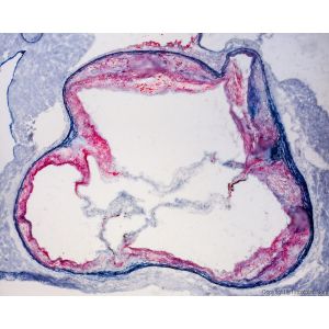
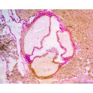
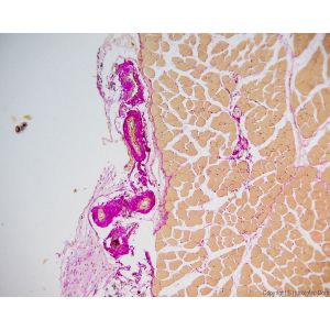
The Hito Verhoeff OptimStain™ Kit is designed based on the Van Gieson and Verhoeff staining method with improved and simplified procedures. This kit enables simultaneous visualization of collagen and elastic fibers.
One key application is in atherosclerosis research. Atherosclerotic plaques contain lipids, inflammatory cells, smooth muscle cells, connective tissue (collagen, glycosaminoglycans, elastic fibers), thrombi, and calcium deposits. Fibrillar collagen is a critical component of lesions: uncontrolled accumulation leads to arterial stenosis, while excessive breakdown weakens plaques, making them prone to rupture.
With its special design, Hito Verhoeff OptimStain™ Kit allows localization and visualization of collagen and elastic fiber damage, providing valuable insights into the extent of atherosclerosis. It can also be used to demonstrate changes in elastic tissues in cases of emphysema and other vascular diseases.
Delivered in a ready-to-use format, the kit ensures high-quality, reliable, and sensitive staining. It has been extensively tested on hearts and arteries from several animal species, offering a simple and effective solution for histological research.
Availability:
In stock
SKU
HTKCS0101
Hito Verhoeff OptimStain™ Kit is designed based on the Van Gieson and Verhoeff staining method with improved and simplified procedures. This kit can be used for simultaneously demonstrating the morphological details of collagen and elastic fibers.
One example application of this kit is to determine the extent of atherosclerosis. The atherosclerotic plaque contains lipids, inflammatory cells, smooth muscle cells, connective tissue (eg, collagen, glycosaminoglycans, elastic fibers), thrombi, and calcium deposits. Fibrillar collagen is a critical component of atherosclerotic lesions. Uncontrolled collagen accumulation leads to arterial stenosis, while excessive collagen breakdown combined with inadequate synthesis weakens plaques thereby making them prone to rupture. With its special design, Hito Verhoeff OptimStain™ Kit will allow localization and visualization of the collagen and damage of elastic structure, therefore, enables a good understanding of the extent of atherosclerosis. This kit can also be used to demonstrate changes in elastic tissues in case of emphysema and other vascular diseases.
Hito Verhoeff OptimStain™ Kit is made in a ready-to-use format and provides high quality, reliable and sensitive staining of collagen and elastic fibers.
Hito Verhoeff OptimStain™ Kit has been tested extensively on the hearts and arteries from several species of animals and it is a simple solution for your research.
| Kit Contents (for >150 slides) | |
| Solution-1A | 125 ml |
| Solution-1B | 55 ml |
| Solution-1C | 55 ml |
| Solution-2 | 225 ml |
| Solution-3 | 225 ml |
| Solution-4 | 225 ml |
| Solution-5 | 225 ml |
| Staining jars | 5 |
| User Manual and MSDS | 1 |
|
Before using Hito Verhoeff OptimStain™ Kit, please make sure you have the following Required Equipment / Materials in your lab (not included in the kit): Cryostat or Microtome, Light microscope |
|

Write Your Own Review
Check items to add to the cart or